What is a blistered cup fungus?
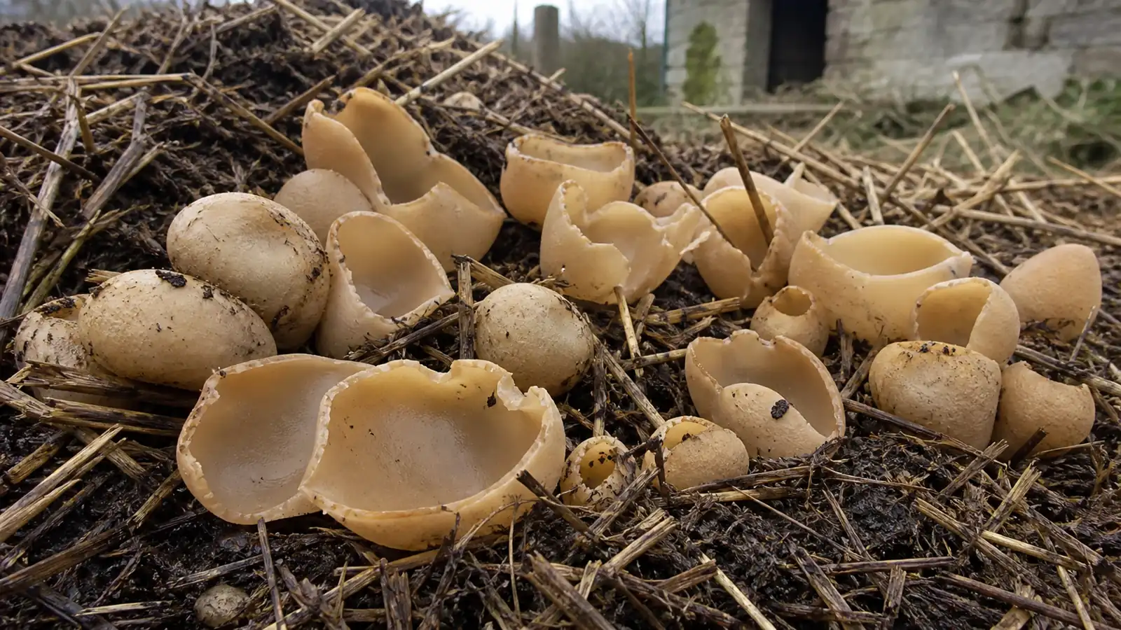
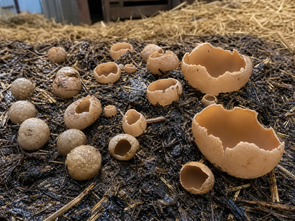
Blistered cup begins as a rounded pale bladder pushing through wet straw or compost. The top opens and expands into an irregular bowl whose smooth tan interior contrasts with a paler, folded, or blistered outer surface.
Crowded growth distorts the cups. Neighbors press against one another, producing lobed mouths and tall compressed walls that look different from a solitary specimen in a field guide.
Photograph the colony as a developmental sequence. Closed young bladders and newly opened cups communicate P. vesiculosa better than one old flattened bowl.
There is no ordinary cap, stem, or gill arrangement.
The inner surface is the spore-producing hymenium, where microscopic sacs called asci develop and release ascospores.
Mature asci can discharge together when humidity changes or the cup is disturbed, creating a visible puff above a colony. This event is spore release, not smoke, steam, or a defensive toxin cloud.
The flesh is brittle and easily splits when a wet cup is lifted.
Handle a scientific voucher from beneath with some attached substrate rather than pinching the rim and losing the base.
Color ranges from cream and buff to tan or ocher. Sun, rain, manure staining, and age can darken the surface, so color names should be recorded under natural light rather than used as the sole identification.
The common names bladder cup and blistered cup describe shape, while common dung cup describes ecology. Taken together they give a useful search image, but none is exclusive to one species.
For most readers the practical questions are whether the fungus harms a compost pile, whether it is safe to eat, and whether an indoor appearance signals moisture.
It is a decomposer, not food, and the substrate issue matters more than eliminating every outdoor cup.
How do you identify a brown Peziza cup?
No separate cap. The fruiting body begins as a pale closed bladder and expands into an irregular tan to ocher cup 3 to 10 cm wide with a blistered or folded exterior.
No gills or pores. The smooth inner cup is the fertile hymenial surface containing asci.
Usually absent or reduced to a short narrowed attachment hidden in manure, straw, compost, or rich soil.
Brittle, pale, waxy flesh without a distinctive pleasant food odor.
Start with the whole colony. Photograph the manure, straw, compost, mulch, or enriched soil and show whether young closed cups occur beside mature open ones.
Cut one mature fruiting body vertically from rim through attachment. Record wall thickness, color layers, brittleness, and any latex or color change, since unexpected features may redirect the specimen.
- Document substrate, management history, moisture, and the full colony.
- Select young, intermediate, and mature stages when collection is permitted.
- Make a clean cross-section and dry specimens promptly at gentle heat.
- Examine mature spores, asci, paraphyses, and excipulum tissue with a Peziza key.

The white spore mass is difficult to use like a gilled-mushroom print because spores launch from the upright inner cup.
A dark glass slide or paper positioned over and beside mature cups may capture a deposit, but microscopic maturity matters more than its pale color.
Spores should be measured from mature material. Immature spores inside asci can be smaller, lack final ornamentation or oil droplets, and lead a novice toward the wrong key couplet.
Paraphyses are sterile filaments among the asci. Their tip shape, pigmentation, and contents can matter alongside the spores, which is why a photograph-only identification of a brown Peziza often stops at genus.
The excipulum, or cup wall, contains tissue layers that can be studied in a thin section. A crushed mount loses the arrangement, so a clean radial slice is more informative than scraping the inner surface.

Record whether the colony is outdoors, in a greenhouse, barn, cellar, or occupied room.
That setting changes the practical response even when the taxonomic procedure remains the same.
For an outdoor compost colony, the fungus can be left to decompose material. For an indoor colony, photograph it, avoid aerosolizing dry debris, and investigate the leak or condensation that keeps the substrate wet.
A species record should retain photographs of every stage, a dried voucher, collection date, substrate composition, and microscopic measurements.
Cup fungi are particularly likely to be renamed when an old record contains only a top-view image.
Measure cup width, height, wall thickness, and attachment across young and mature material. Crowding changes outline, so one compressed cup cannot define the normal shape range.
The inner hymenium should be sampled from a mature undamaged area. Edge tissue may contain immature asci, while the oldest center can be contaminated or discharged, making several small mounts more useful than one scrape.
Use water, iodine reagent, cotton blue, or another mount only as required by the selected Peziza key. Reagent responses are interpretable when preparation and concentration are recorded, not when a color is remembered afterward.
Substrate belongs on the specimen label. Preserve a small dry sample or detailed description of manure, straw, wood fiber, soil, and indoor material because later taxonomy may depend on ecology.
If a culture is obtained, link its number to the fruiting voucher. A clean mycelial plate without the original cup and its microscopy cannot independently prove P. vesiculosa.
Which cup fungi share the same shape?
Brown and tan Peziza cups overlap strongly in shape. The blistered cup is suggested by its crowded pale cups on dung-rich straw or manure, but microscopy and substrate records are needed for a confident species determination.
Do not eat a brown cup fungus. Macroscopic features overlap across Peziza, and the manure-rich habitat creates a separate contamination risk even when the species name is correct.
| Candidate | Typical setting | Character that changes the comparison |
|---|---|---|
| Peziza vesiculosa | Manure, dungy straw, compost, silage residue | Crowded bladder-like young cups and blistered exterior |
| Peziza varia group | Soil, mulch, wood debris, buildings | Layered flesh under microscopy and a broader range of substrates |
| Peziza repanda | Bare soil and wood-rich ground | Broad spreading cups with less bladder-like clustered development |
| Peziza domiciliana | Damp buildings, mortar, carpet, or construction material | Indoor substrate and moisture problem |
| Otidea species | Woodland litter and soil | Ear-shaped or split fruiting bodies rather than closed young bladders |
The substrate is evidence, not proof. A tan cup in compost may be another Peziza, while P. vesiculosa can fruit where manure has been mixed so thoroughly that no obvious dung remains.
Young fruiting bodies matter because the common name describes their inflated, blister-like form. Old cups flatten, tear, and resemble many other brown cup fungi after rain.
A cross-section should show brittle homogeneous flesh, but species-level Peziza work often needs microscopic tissue layers, mature spores, asci, and paraphyses. A photograph of the upper surface alone cannot provide them.
Brown cup shapeDozens of cup fungi converge on tan or ocher bowls.
Compost locationEnriched substrate supports several saprobes.
Developmental seriesPhotograph closed bladders, opening cups, and mature torn fruiting bodies together.
Microscopic voucherMature asci and spores let a specialist evaluate the name.
Indoor cups deserve a building-moisture response rather than a food or pesticide experiment. Find and correct the wet material, then remove contaminated porous substrate under appropriate mold-safety practices.
Why does it grow on manure and wet straw?
Peziza vesiculosa is a saprobe that uses nutrient-rich decomposing organic matter. Manure, urine-enriched bedding, wet straw, silage residue, and compost provide nitrogen and water beyond ordinary woodland litter.
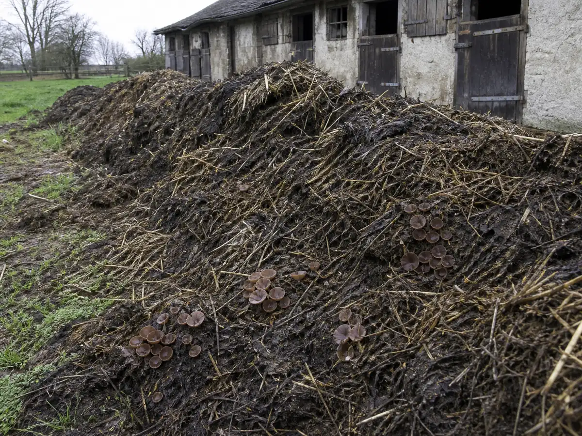
The fungus helps break down plant fibers and animal-waste mixtures. Its presence outdoors does not mean the compost is ruined, although it can indicate a wet, cool, locally compacted layer.
Fruiting converts part of the substrate into fungal biomass and airborne spores while the hidden mycelium continues digestion.
Warning
Persistent indoor cups indicate enough moisture for fungal growth. Find the water source instead of treating the visible cups as the whole problem.
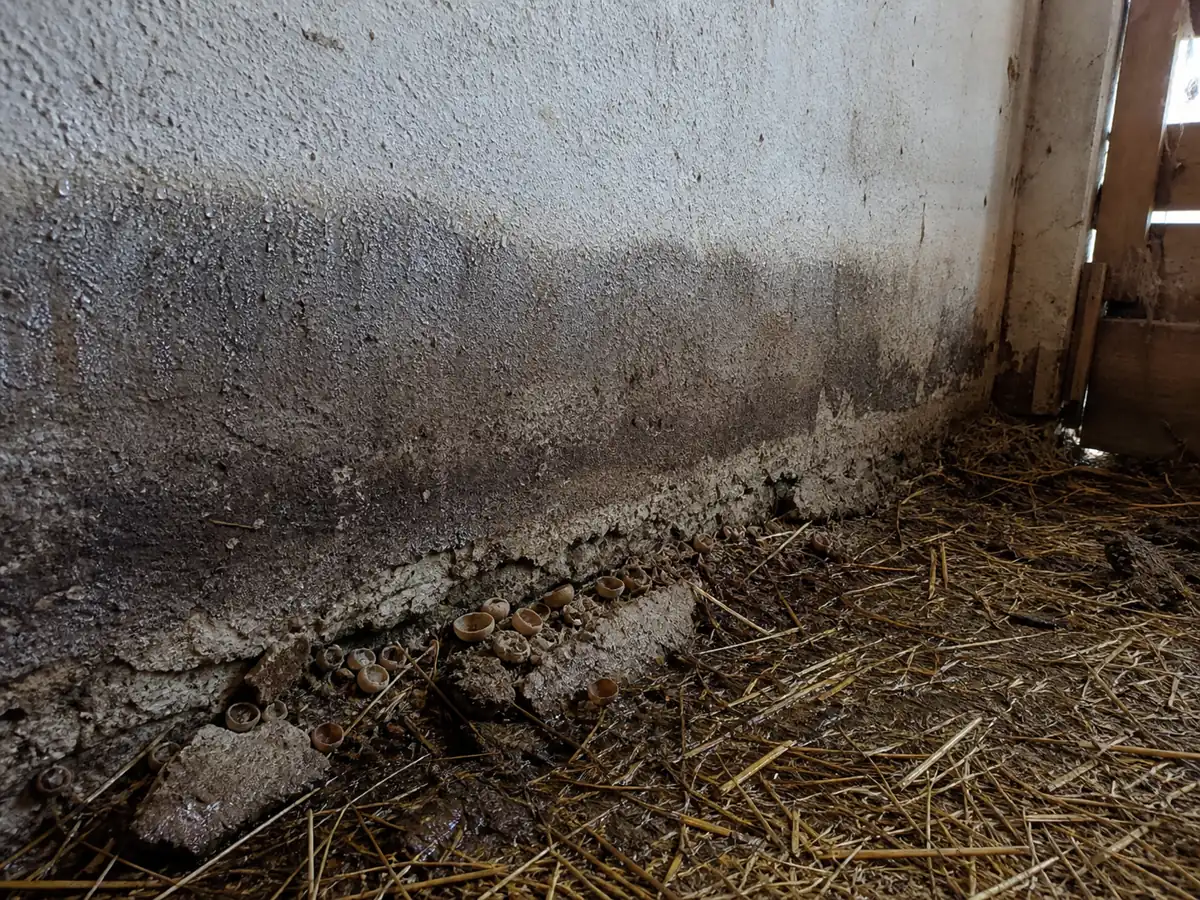
A hot active compost core may be too warm for fruiting, while cooler edges and aging piles allow cups to form. Turning the pile changes oxygen, moisture, temperature, and the physical colony without requiring fungicide.
Agricultural sites can create repeated flushes as new bedding or manure is added.
Record whether the cups sit on fresh material, a weathered surface, or buried older straw because that detail helps interpret the substrate.
Spore puffs distribute microscopic propagules, but a visible colony usually reflects suitable wet material already present rather than a sudden invasion from the latest puff. Drying or removing the substrate limits fruiting more effectively than spraying the cups.
Outdoor fruiting bodies feed invertebrates and eventually collapse back into the pile. There is no need to remove them unless people, livestock management, or a scientific voucher creates a specific reason.
Indoor fruiting changes the decision because drywall paper, carpet backing, wood fiber, or accumulated organic debris may remain chronically damp. Fixing the source of moisture helps prevent recurrence and protects the building better than removing the visible cups alone.
Livestock access changes the safety context around a colony.
Keep children and pets away from manure piles, and follow farm hygiene rather than treating the cup fungus as the only biological hazard.
Compost temperature should be measured below the surface because fruiting at a cool edge says little about the core. A pile can support cups in one wet pocket while decomposing normally elsewhere.
In a greenhouse, repeated cups may point to overwatering, poor drainage, and a reservoir of old organic media. Adjust those conditions for plant and worker health instead of applying a fungicide to each fruiting.
The wet substrate owns the recurrence. Remove or rebalance that material and fruiting declines, while cutting cups alone leaves the mycelium and moisture untouched.
Outdoor spores are normal and widely dispersed. The practical control is unsuitable indoor moisture, not an attempt to sterilize open air or a working compost system.
Why does microscopy matter for Peziza?
Peziza vesiculosa was described by Jean Baptiste François Bulliard and remains in Pezizaceae. Historical combinations placed it in genera such as Aleuria, Galactinia, Plicaria, Pustularia, and Scodellina.
Those synonyms reflect changing classifications of cup fungi rather than multiple ordinary household species. Current databases accept P. vesiculosa, but local records still need microscopic confirmation because Peziza morphology is convergent.
- Ascomycete
- Spores mature inside asci on the inner cup rather than on basidia covering gills.
- Apothecium
- The entire open cup is a fruiting structure that exposes the hymenium to air.
- Species limit
- Macroscopic blisters and manure habitat suggest the name but do not replace mature-spore and tissue study.
DNA data can resolve collections that remain ambiguous after microscopy, especially within groups where spore size and cup color overlap. Sequencing is most useful when linked to a well-documented voucher rather than an environmental sample with no fruiting morphology.
Should you remove it from compost?
There is no good reason to cultivate blistered cup as a household mushroom. It fruits opportunistically on rich wet organic debris, but a deliberate manure culture would create sanitation and spore-management problems without food value.
If it appears in compost, adjust the pile only for composting goals. Balance moisture, aeration, carbon-rich material, and temperature rather than applying fungicide to one decomposer.
If it appears indoors, do not encourage or transplant it. Identify and repair the moisture source, remove damaged porous material safely, and dry the assembly.
A sudden outdoor flush often follows a local moisture pocket rather than failure of the whole pile. Push a gloved hand or compost thermometer into several zones and compare water, heat, and compaction before changing the recipe.
Adding dry shredded leaves, clean straw, or another suitable carbon source can improve structure when the pile is sodden and nitrogen-rich. The goal is aerobic decomposition, not the elimination of every fungal fruiting body.
Indoor growth calls for finding and correcting the source of moisture. Inspect plumbing, roof entry, condensation, slab edges, and ventilation, then verify that the affected assembly dries after repair.
Do not sweep a mature dry colony in an occupied room. Lightly contain the material, use appropriate protective equipment, and follow local guidance when contamination covers a large area or involves sewage.
Can you eat blistered cup?
Do not cook or taste blistered cup. Even if a field guide reports historical use for a brown cup fungus, visual overlap and contaminated substrate make it an unsuitable food experiment.
Washing cannot remove uncertainty about species or manure-associated microbes. Heat also does not restore the missing identification evidence in chopped cups.
Keep it out of mixed wild-mushroom baskets. Brittle fragments lose the cup wall, attachment, and substrate clues that a specialist would need after an accidental collection.
Blistered cup and compost questions
Is blistered cup edible?
Why is it called blistered cup?
Does it damage compost?
Why can a cup release a puff?
Can it grow indoors?
Can a photograph confirm Peziza vesiculosa?
Sources & References
- NatureServe Explorer Accepted taxon and common-name record based on Species Fungorum.
- GBIF Backbone Taxonomy Accepted name and historical combinations.
- Burke Herbarium Image Collection Voucher imagery and North American name context.
- British Mycological Society Cup-fungus recording and microscopy context.