What makes this webcap blood red?
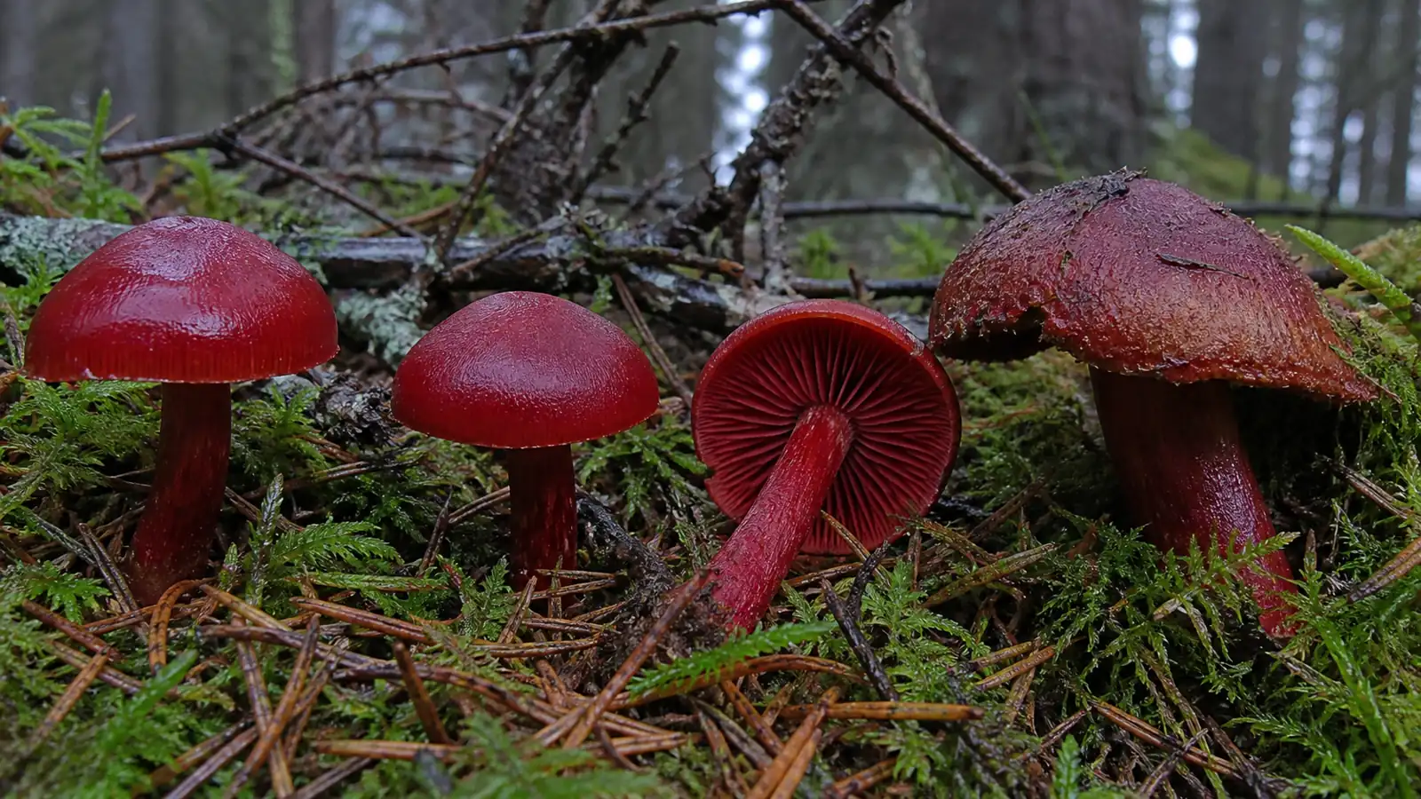
Blood-red webcap earns its name through an unusually complete red color scheme. A fresh fruiting can show dark red cap fibers, scarlet to blood-red gills, a red stem, and red flesh in one small mushroom.
As the spores mature, rusty dust softens the gill color and collects on the stem where the cortina once stretched. This aging sequence can make an old specimen look much browner than a young one.
Document the mushroom before and after the rusty spores appear. Young veil and gill colors explain the identity better than a single mature cap.
The cap surface is dry and finely fibrillose rather than slimy or waxy. Its color may be deepest at the center and fade toward a margin that remains curved downward while young.
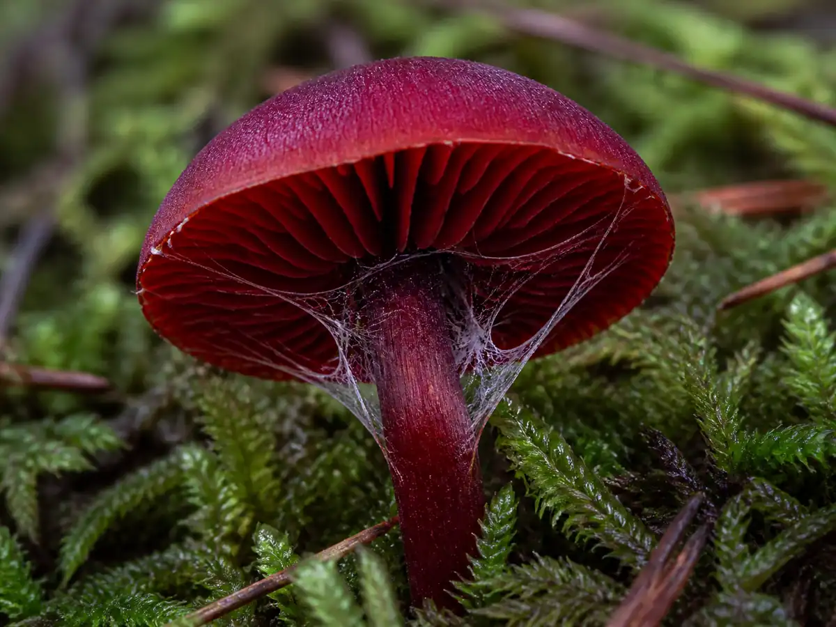
The cortina is not a solid skirt.
It consists of delicate radial fibers, and a clean side photograph of an unopened specimen is often the only permanent evidence that it existed.
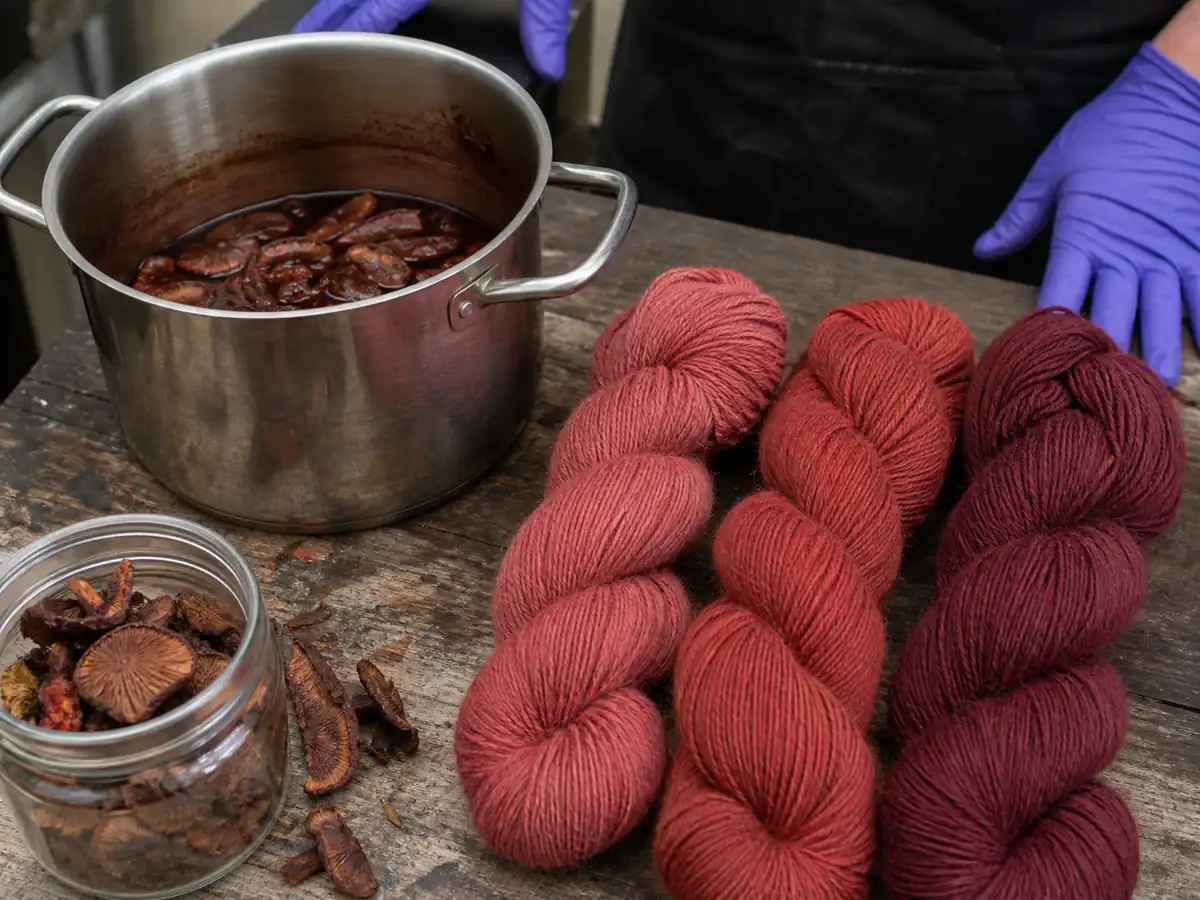
The blood-red pigments include anthraquinone compounds studied for dyeing. A strong textile color does not establish food or medicinal safety, and extraction can concentrate chemicals that have not been evaluated as household products.
Common-name searches often mix C. sanguineus with related Dermocybe-type webcaps.
The visible all-red pattern is valuable, yet region and sequence evidence decide whether a record belongs to the European species or a related lineage.
Distinguish the full-red form from webcaps with red gills and a differently colored cap. Preserve evidence after suspected exposure, and do not assume that every red Cortinarius contains the same toxin.
A conservative response does not require knowing the exact molecule in the specimen. The combination of genus uncertainty, poisonous status, and dangerous relatives is enough to keep it out of food.
How do you document the cortina and red flesh?
Convex to broadly domed, usually 2 to 5 cm, dry and finely fibrillose, colored deep blood-red to red-brown and darkening with age.
Crowded, attached, and vivid blood-red when young, becoming rust-stained as brown spores mature.
Slender, red to dark red-brown, 3 to 8 cm long, with yellowish basal mycelium and rusty remnants of a cobwebby cortina rather than a solid ring.
Red to dark red flesh with a mild or radish-like odor; do not taste any webcap.
Begin with young fruiting bodies. Photograph the intact cortina, cap texture, and bright gills before air, fingers, and spores change their appearance.

Produce a print from a mature cap on pale material. The rusty-brown deposit supports Cortinarius but is shared across the genus and cannot separate poisonous species.

Preserve the cortina
Photograph unopened cortina fibers from the side.
Map fresh color
Record fresh colors of cap, young gills, stem, base, and cut flesh.
Record the host setting
Note spruce, pine, moss, soil moisture, and neighboring trees.
Retain a voucher
Dry a complete voucher for microscopy or sequencing when collection is lawful.
The cap should be examined for radial fibrils under diffuse light.
A smooth waxy red cap with thick waxy gills and a white print redirects the collection toward Hygrocybe rather than Cortinarius.
Cut one specimen lengthwise. C. sanguineus carries red into the flesh, while several related webcaps have paler internal tissue or concentrate stronger color in the gills.
Cortina remnants may look rusty after catching spores. Separate the original veil color from later spore staining by examining a protected young stem and the cap-margin attachment.
Microscopy evaluates roughened brown spores, their size and shape, cystidial structures where relevant, and cap tissues. Pigment reactions and DNA can become important in Dermocybe-type groups where macroscopic colors overlap.
Do not wash a voucher.
Water removes spore dust, mats cap fibrils, strips cortina fibers, and blends soil into the basal mycelium.
Place each stage in a separate breathable container with its field number.
Color calibration matters for red fungi. Include a neutral gray reference in one field photograph and avoid heavy phone filters that push brown-orange mushrooms toward scarlet.
Record changes during drying because intense reds can darken or fade. The dried specimen should remain linked to fresh photographs, notes, and microscopy so later reviewers do not reconstruct the color from memory.
For a poison exposure, the identification standard changes from a perfect species record to rapid evidence preservation. Save uncooked specimens, meal remnants, photographs, collection location, time eaten, and symptom timeline for the poison center and treating team.
Pigment extraction is not part of a field identification, but a specialist may record color reactions in cap, gill, and veil tissue with standardized reagents.
The specimen and method must remain linked because free-form descriptions such as turned darker are not repeatable evidence.
Roughened spores should be measured in side and face view. Shape can appear different when a spore rolls, so a representative series is more reliable than one ideal outline.
The young gill color deserves protected material. Place one unopened fruiting body in its own container so red gills and unstained cortina remain separate from rusty spores shed by mature caps.
Preserve the color map before the print. Once rusty spores cover the gills and veil, the observer can no longer tell which brown-red zones belonged to fresh tissue.
For a difficult regional record, sequence data should come from the photographed voucher. An environmental root sequence or an unlabeled red cap cannot establish the same evidence chain.
If someone may have eaten this mushroom, act now. Call Poison Control at 1-800-222-1222 (US) or emergency services before symptoms appear, and save a piece of the mushroom for identification.
Which red webcaps can be confused with it?
Red webcaps form a difficult Cortinarius group. The blood-red cap, gills, stem, and flesh support C. sanguineus, while several relatives carry red only on the gills or combine it with olive, yellow, or brown surfaces.
Warning
Never eat a red or brown webcap. The genus contains dangerously poisonous species, including mushrooms associated with delayed kidney injury, and field characters do not provide an edible shortcut.
| Candidate | Distribution of red color | Useful contrast |
|---|---|---|
| Cortinarius sanguineus | Cap, young gills, stem, and flesh are strongly red | Mossy conifer habitat and blood-red overall appearance |
| Cortinarius semisanguineus | Young gills are blood-red | Cap is olive-brown to yellow-brown rather than fully red |
| Cortinarius cinnabarinus | Cinnabar to orange-red fruiting body | Often associated with deciduous woodland and different microscopic characters |
| Cortinarius phoeniceus group | Red gills with brown or olive cap tones | Variable veil pigments, ecology, and DNA |
| Hygrocybe species | Bright red cap and gills in grassland | Waxy texture, white spores, and no rusty cortina |
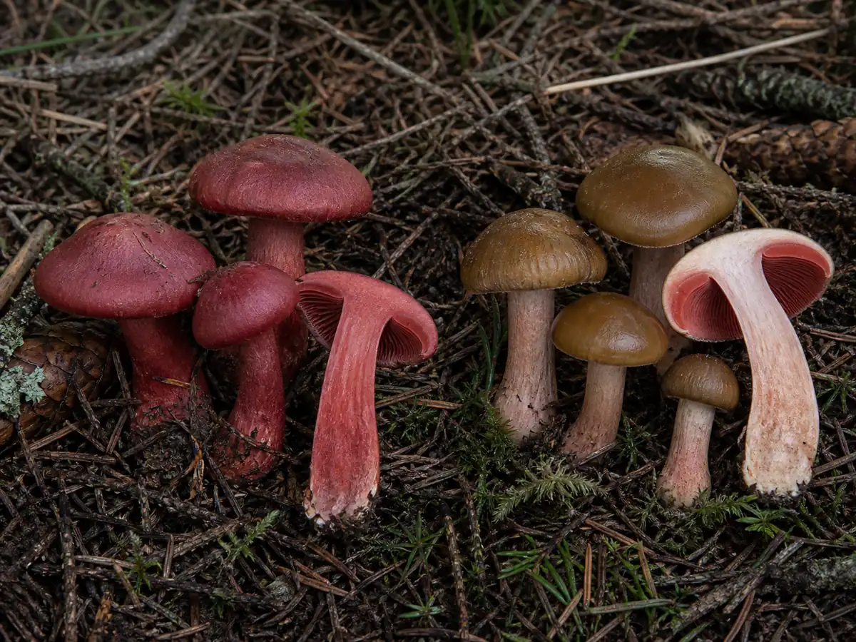
The cortina is most visible on young unopened caps as fine fibers stretching between cap edge and stem. It collapses quickly and later catches rusty spores, leaving a faint fibrillose zone rather than a membranous ring.
C. semisanguineus is called half-blood-red because the vivid color is concentrated in the gills. A brown or olive cap above red gills should reopen that comparison rather than be explained as weathering.
Old C. sanguineus can look brown-red after rain and spore deposition. Cut flesh and protected young gills preserve red evidence longer, but microscopy and a current regional key remain necessary for a scientific record.
A missing or damaged stem base should stop a food or species claim. Basal mycelium, veil remnants, and color can be lost when a collector snaps the mushroom at ground level.
Red brittlegills can share a woodland color but break with a chalk-like snap, lack a cortina, and produce a pale print. Their presence in the same moss makes texture and veil evidence more useful than a distant red-cap photograph.
Red waxcaps usually occupy grassland and have thick waxy gills with a white print. A woodland edge can place turf and trees close together, so substrate must be shown rather than inferred from the background.
Some red webcaps are rare enough that destructive comparison should be limited.
Photograph a developmental series and collect only the material a lawful voucher requires.
No red Cortinarius is a casual food candidate. Even when the exact species remains unresolved, the genus-level evidence already answers the eating question.
Why does it fruit in mossy conifer woods?
Cortinarius sanguineus forms ectomycorrhizae with living trees. It is strongly associated with conifer woodland in European accounts, often appearing among moss beneath spruce or pine.
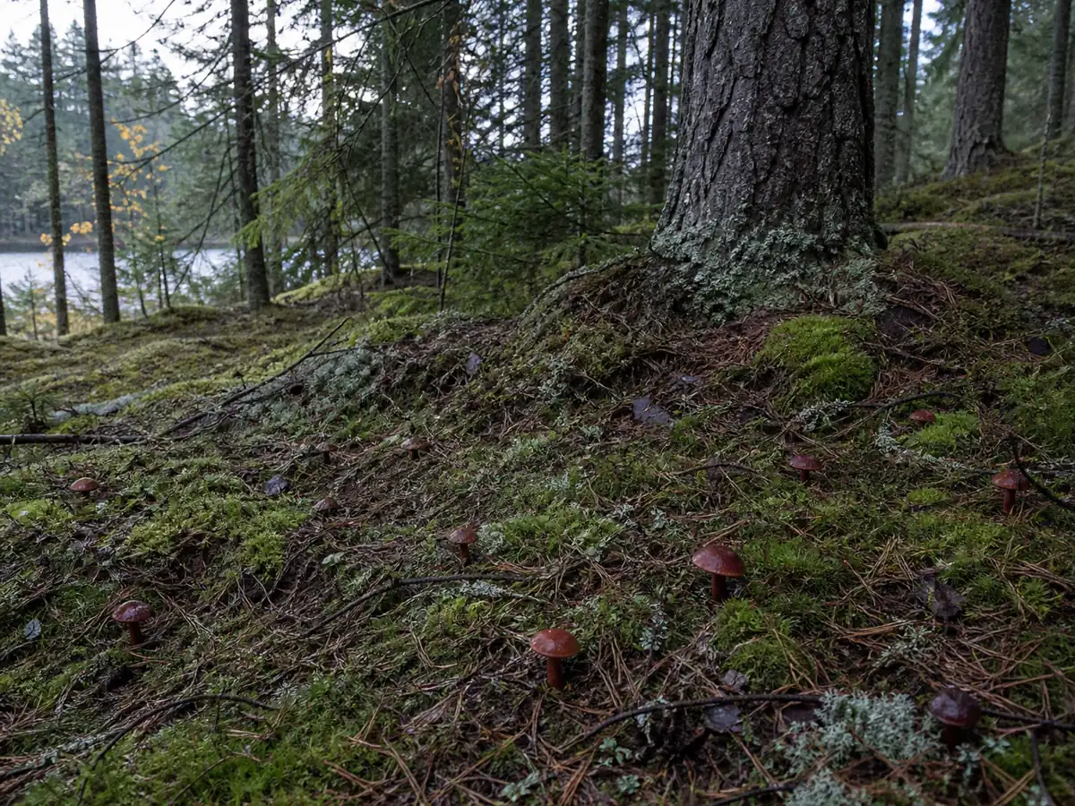
The mycelium surrounds fine roots and extends into soil, receiving plant carbon while improving the root system's access to water and mineral nutrients. The red fruiting body is a temporary reproductive structure of that persistent partnership.
Moist moss buffers temperature and water around the developing caps. Dark shaded stands can preserve the saturated red longer than exposed edges where sun and drying shift fruiting bodies toward brown-red.
Note
The nearest trunk is evidence, not proof of the host. Conifer roots cross beneath moss, and a mixed stand may connect the fungus to a tree outside the photograph.
Record every plausible host, soil type, drainage, moss cover, slope, and recent rainfall.
Those facts help separate ecological patterns among visually similar webcaps.
Fruiting commonly occurs from late summer into autumn after adequate moisture. A known site may skip dry years while the underground fungus remains alive with its host.
Do not rake moss to find more specimens. Disturbance damages fine roots and microhabitat, while one complete permitted voucher is usually more useful than a bag of broken red caps.
The pigments persist after collection and have attracted dye research, but wild harvest for color can remove a scarce regional fruiting. Conservation status and land permission should be checked before collecting for any purpose.
Where does Dermocybe fit in its name?
Cortinarius sanguineus belongs to the red-pigmented group historically treated as Dermocybe. Some classifications raised Dermocybe to genus level, while broader treatments retained these species in Cortinarius.
Modern phylogenetic work has reorganized Cortinarius and related genera, and accepted combinations can differ between databases. The scientific concept should therefore be tied to a cited treatment and region rather than inferred from the word webcap.
- Name meaning
- Sanguineus refers to the blood-red color spread across the fruiting body.
- Comparison
- Semisanguineus means half-blood-red and describes a relative with red gills beneath a browner cap.
- Pigment group
- Anthraquinones help explain the strong dye colors but do not define edibility.
Older poison and dye literature may use Dermocybe sanguinea or a different genus boundary. Search historical combinations when reviewing chemistry, then check that the material matches the modern species concept.
Sequence-backed vouchers are important outside northern Europe because visually similar red webcaps occur elsewhere.
A global image search can join unrelated lineages into a false worldwide distribution.
The historical subgenus or genus Dermocybe grouped webcaps whose yellow, orange, and red anthraquinone pigments were conspicuous. Molecular classifications have tested and rearranged those boundaries rather than preserving color as the only relationship signal.
Chemical studies should cite the accepted voucher name and any historical combination. Dermocybe sanguinea in an older dye paper may refer to the same intended species concept, but that connection needs confirmation.
Pigment similarity is not toxin identity. Red relatives can share dye compounds while differing in other chemistry, and no color lets a forager infer a safe meal.
Distribution records outside Europe deserve sequence-backed review because the same saturated red pattern has evolved or persisted in related regional lineages. A location tag and photograph alone do not establish a global C. sanguineus range.
For public databases, note the taxonomic authority and determination method. That transparency lets future reviewers update the name without losing the observation.
Do its dye pigments have medical value?
Laboratory studies of C. sanguineus pigments address colorants and cell toxicity, not clinical treatment. An extract that dyes fiber or affects cultured cells has not demonstrated a safe human dose or therapeutic benefit.
Do not make a tincture or topical preparation from a poisonous webcap. Skin exposure to dyes, solvents, and concentrated extracts creates a different risk from ordinary field handling.
Use gloves for intentional pigment work, avoid food equipment, label all material, and follow a researched dye protocol only where collection is legal. These controls support chemical craft, not medicine.
Can blood-red webcap be eaten?
There is no culinary preparation for blood-red webcap. Do not taste, parboil, pickle, dry, or mix it with edible woodland mushrooms.
The red gills can resemble a dramatic chanterelle to an inexperienced collector, but chanterelles do not carry a rusty spore print or cobwebby cortina. A remembered color is not a safe substitution for structure.
After accidental mixing, discard the affected basket as food and retain separate evidence for identification. Tiny fragments coated in rusty spores cannot be sorted reliably in a pan.
Blood-red webcap safety questions
Is blood-red webcap edible?
What color is its spore print?
What is a cortina?
How is it different from Cortinarius semisanguineus?
Does red color prove orellanine?
What should happen after ingestion?
Sources & References
- First Nature field account European morphology, habitat, comparison, and conservative toxicity context.
- Waterless Dyeing and In Vitro Toxicological Properties Identified anthraquinone pigments and limits of available toxicology.
- GBIF Backbone Taxonomy Taxonomic and distribution record.
- British Mycological Society Regional recording and Cortinarius field context.