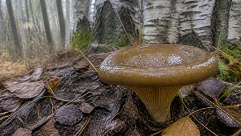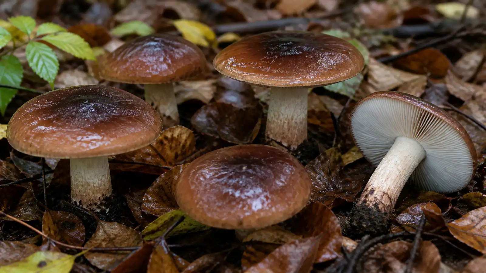
What makes bitter poisonpie more than another brown cap?
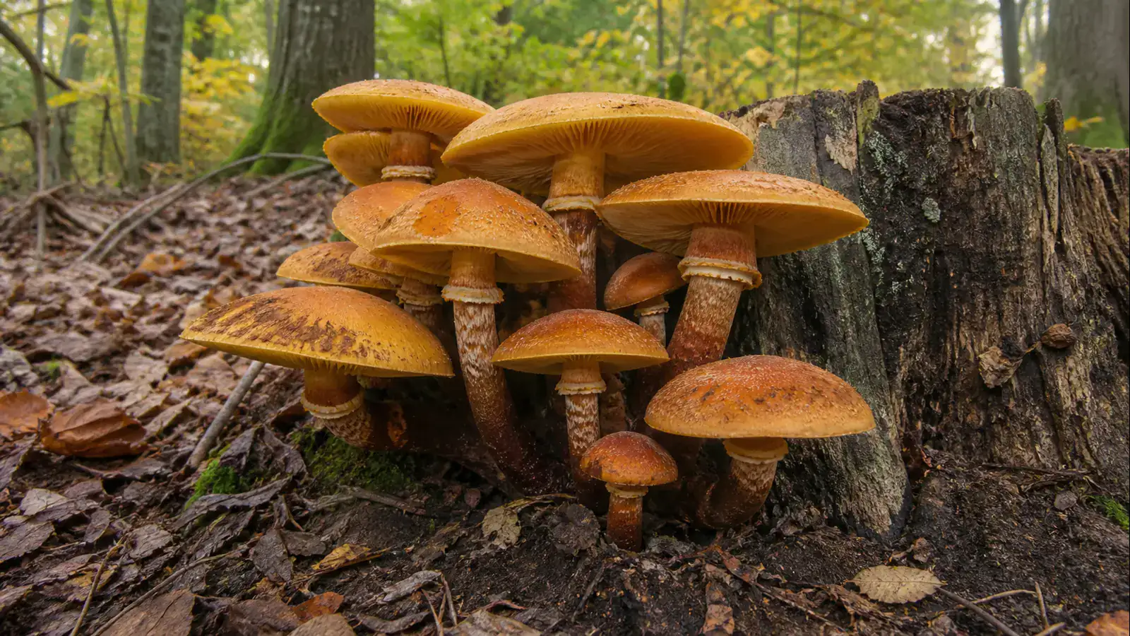
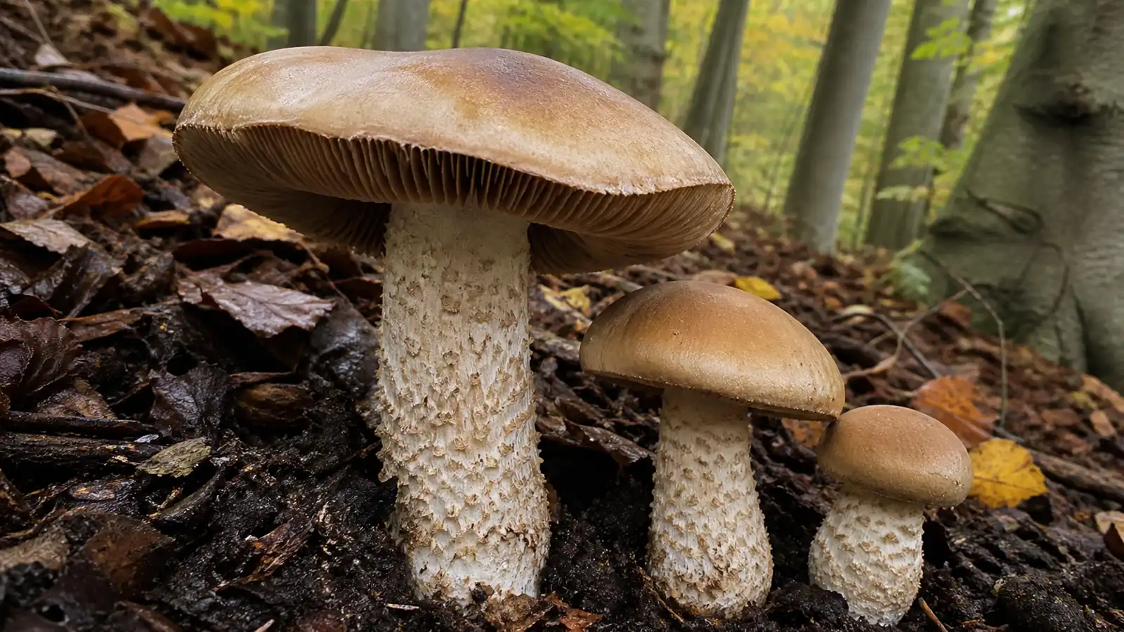
Hebeloma sinapizans is one of the larger poisonpies. Its broad ochre or clay-colored cap stands above a thick pale stem whose coarse scales can look dusted or shaggy.
The gills begin pale and darken to cinnamon or rusty brown. A radish-like smell fits the genus but does not separate every species.
Cut the mushroom lengthwise for documentation. In mature specimens, a triangular tongue of cap flesh descends into the hollow top of the stem, a feature traditionally used to distinguish this species.
Key takeaway
Large size and a rough stem narrow the field, but safe handling still requires a brown spore print, the internal stem-apex character, habitat, and microscopic confirmation where needed.
The stem-apex projection is easiest to see in a mature specimen cut cleanly from cap center through the full stem. It is a useful discriminator inside the Hebeloma complex, but a damaged, immature, or waterlogged mushroom may not show it clearly.
The common name little brown mushroom understates the challenge. Odor, cap tackiness, gill droplets, stem ornament, spore color, microscopic structures, host, and geography can all change which Hebeloma name is defensible.
This is not always a tiny brown mushroom. Mature caps can be broad and substantial above a thick coarsely textured stem, which makes the species look more reassuring than the little-brown-mushroom warning suggests.
The radish-like odor, clay-to-cinnamon gills, ringless scaly stem, rusty print, and growth with living tree roots create a useful genus-level pattern.
None of them alone separates every Hebeloma, and the poisonous food decision remains easier than the species name.
Cutting the mushroom lengthwise can expose a distinctive projection of cap flesh that extends into the hollowing stem apex. That feature is valuable when present, but age, damage, and related species can weaken its appearance.
Hebeloma taxonomy combines morphology, ecology, microscopy, and DNA because older broad names grouped several lineages together. A large brown cap near beech may support H. sinapizans while still requiring spores and gill-edge cells for a defensible record.
Does the cut stem reveal the decisive clue?
Clay-buff to ochre-brown, 5 to 15 cm, convex then broadly expanded, often greasy when wet.
Crowded, notched, pale clay then cinnamon-brown as spores mature; edges may bead with moisture.
Thick, whitish to buff, without a ring, covered in coarse scales or fibrils, often with a swollen base.
Thick whitish flesh with a radish-like odor and bitter taste; a wedge of cap flesh projects into the hollow stem apex.
Record fresh cap texture, gill attachment, stem scales, odor, and the intact base before cutting. Wear gloves if handling will be prolonged and wash hands afterward.

Hebeloma crustuliniforme is generally smaller and lacks the same pronounced internal projection, but field overlap is substantial. Cortinarius can share rusty spores yet often shows a cobwebby veil or rusty cortina remnants.

Use the spore print inventory to compare this rusty deposit with other print-color groups. Species-level Hebeloma work still depends on spore ornamentation, cheilocystidia, ecology, and DNA, so a phone photograph is not enough for a food decision.
Document before the specimen dries because a tacky cap can become dull and the radish odor can weaken. A field note should separate what was observed fresh from what appeared later under indoor light.
Under magnification, specialists compare spore size and ornamentation with cheilocystidia along the gill edge. Those characters are especially important when the cap-flesh projection is weak or a regional key includes several large scaly-stemmed species.
Rusty spores rule out white-spored lookalikes, but they also keep Cortinarius and many other brown-spored mushrooms in play.
Check for veil remnants, gill attachment, stem interior, and the complete base before narrowing the genus.
Collecting one young, one mature, and one old specimen from the same flush can show whether gill color, stem hollowing, and cap texture form a coherent developmental series. Keep the bases intact and label specimens by location so mixed brown mushrooms are not combined accidentally.
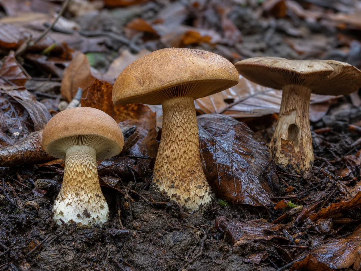
Photograph cap texture before it dries indoors. A moist surface can feel tacky and look slightly glossy, then become dull, paler, or cracked, which changes the character that a later observer sees.
Smell a freshly cut section without tasting it. The radish or mustard-root note may be strongest in young firm tissue and can fade in old waterlogged specimens, so a weak odor is missing evidence rather than an edible signal.
The gills begin pale clay and mature toward cinnamon as spores accumulate. Note the notched attachment, edge texture, and any droplets or spotting before making a print, since print color alone leaves many Hebeloma and Cortinarius candidates.
Cut from the cap center through the entire base.
Record the internal cap-flesh projection, hollowing, flesh color, stem scales, and absence of a true ring in one image so the clue is not detached from the rest of the specimen.
Under a microscope, experts compare spore dimensions and ornamentation with cheilocystidia on the gill edge. DNA and a regional key become important when the projection is weak, the host is uncertain, or several large scaly-stemmed poisonpies overlap.
The cap-flesh projection is easiest to evaluate in a clean central cut from a mature but firm specimen. An off-center cut, insect tunnel, or collapsed old stem can create or erase a projection-like shape, so photograph the intact exterior beside the cut half.
| Observation | What it contributes | Why it cannot finish the identification |
|---|---|---|
| Thick ringless stem | Supports the expected robust build | Other Hebeloma share this form |
| Cap-flesh projection | Adds a useful internal character | It may be weak or absent in one specimen |
| Rusty clay-brown print | Rejects white-spored candidates | Cortinarius and other brown-spored fungi remain |
| Radish-like odor | Supports the Hebeloma group | Odor changes with age and handling |
Chemical reagents and microscopy used in specialist keys require controlled interpretation. A color reaction copied from an online table cannot replace a regional description, calibrated measurements, and comparison with authenticated material.
Drying a labeled voucher preserves spores and gill-edge tissue for later review. Keep the site, date, candidate host trees, photographs, and collector attached to that voucher because microscopic measurements without ecology can still match several Hebeloma.
If someone may have eaten this mushroom, act now. Call Poison Control at 1-800-222-1222 (US) or emergency services before symptoms appear, and save a piece of the mushroom for identification.
What should you do after possible bitter poisonpie exposure?
The expected gastrointestinal pattern does not allow self-diagnosis. A meal may contain several wild species, symptoms can come from other causes, and dehydration or severe pain can require medical assessment even when delayed liver toxicity is not expected from H. sinapizans.
Call before discarding the evidence. Photograph raw and cooked material, keep unused specimens cool in a paper container, and record the meal timeline without waiting to see whether every diner reacts.
Do not serve the remainder to test whether the first illness was coincidence. Dose, age, health, and individual response can differ, so another person may have a different outcome from the same dish.
Which brown mushrooms resemble bitter poisonpie?
Hebeloma is a difficult brown-mushroom genus whose species share clay caps, brown spores, and radish-like odors. The distinctive cap-flesh projection helps, but microscopy and a regional key may still be required.
Do not eat any poisonpie or unidentified little brown mushroom. Hebeloma crustuliniforme and related poisonous species overlap, while edible brown-gilled mushrooms must be separated by much more than cap color.
Hebeloma crustuliniforme and related poisonpies can overlap in cap color, odor, and habitat. The internal stem-apex projection supports H. sinapizans only when the specimen is mature and the rest of the character set agrees.
Hebeloma crustuliniforme has been used as a broad name for many poisonpie collections and can share clay-brown color, radish odor, rusty spores, and root-associated habitat. Size and the internal projection can guide comparison, but neither should be treated as a food test.
Cortinarius adds a genus-level hazard. Young specimens may show a cobwebby cortina between cap and stem, and rusty spores later color the gills and veil remnants, but rain or handling can erase that web.
Other brown-spored mushrooms can fruit in the same woodland after rain. Collect one young, one mature, and one old specimen from each apparent group, label them by exact position, and keep every base intact so different developmental series are not merged.
The lookalike question ends at rejection for food. A specialist may continue to microscopy and sequencing, while a cook gains nothing by forcing a name onto an uncertain bitter brown mushroom.
Why must bitter poisonpie stay out of the basket?
Bitter poisonpie is not food.\n\nIts name records the bitter taste, but tasting a poisonous candidate is unnecessary and unsafe.
Cooking does not provide a reliable detoxification method. If it enters a mixed basket, discard the uncertain collection rather than attempting to sort cooked pieces.
Gastrointestinal symptoms can begin after the meal has been cleared away, so preserve a raw specimen and photograph every species in a mixed basket before cooking.
Record the amount, meal time, onset of nausea or diarrhea, and who else ate the same collection for the poison specialist.
The word bitter in the common name is a warning, not an invitation to taste. Deliberately chewing a poisonous candidate adds exposure and cannot distinguish the several brown-spored species that share unpleasant flavors.
No household preparation has been validated to detoxify H. sinapizans. Boiling, soaking, drying, or discarding water also destroys or separates features that poison specialists may need if a meal goes wrong.
Keep wild collections separate until identification is complete. One poisonpie cut into a mixed basket can leave brown cap pieces and gills that are impossible to assign after cooking.
If exposure occurs, nausea, vomiting, cramps, and diarrhea can cause meaningful fluid loss.
Contact a poison service, retain raw and cooked material, record the time and amount, and seek urgent care for persistent vomiting, fainting, confusion, severe weakness, or inability to keep fluids down.
Which tree roots support Hebeloma sinapizans?
Bitter poisonpie forms ectomycorrhizae with living trees and fruits from soil. Beech and oak are frequent partners, with other broadleaf and conifer associations reported.
Fruiting bodies appear singly, scattered, or in groups during late summer and autumn. Parks, pathsides, and disturbed woodland can produce large specimens when suitable roots are present.

The fruiting body is temporary, while the root-associated mycelium remains underground. Do not infer that removing mushrooms removes the fungus or benefits the tree.
Disturbed ground does not mean the fungus is independent of trees. Park lawns, path edges, and planted beds can contain living beech, oak, willow, or conifer roots well beyond the canopy edge.
A flush after rain may produce several age classes at once. Young pale-gilled buttons, mature rusty-gilled caps, and collapsing old specimens can look like separate species unless their shared root zone and developmental sequence are recorded.
Hebeloma sinapizans exchanges resources with living roots rather than decomposing the leaf litter around it.
The cap may rise through thick litter, but the ecological partner lies below ground and can be meters from the nearest visible trunk.
Beech and oak are frequent associates, with other broadleaf and conifer partners recorded in regional treatments. Record every plausible tree, because a mixed park or woodland does not reveal the host from one overhead photograph.
Late-summer and autumn rain can produce a flush with buttons, open caps, and collapsing old mushrooms together.
The change from pale to cinnamon gills and from solid to hollowing stems becomes clearer when those ages are documented as one series.
Managed parks and path edges still contain extensive roots. Mowing or picking caps does not remove the fungus, and disturbing the soil can damage both roots and the mycorrhizal network without solving a safety problem.
Where children or pets may eat the fruiting bodies, remove visible mushrooms and monitor after rain. That exposure control is different from trying to eradicate a root partner from the landscape.
Why are Hebeloma names difficult?
Hebeloma sinapizans belongs to Hymenogastraceae. The genus contains many morphologically similar brown-spored ectomycorrhizal species and has been revised using morphology, ecology, and DNA.
Older broad applications of poisonpie names may hide multiple species. Verified modern records should anchor regional claims.
Modern Hebeloma work combines type material, morphology, ecology, and DNA because older field names were often applied too broadly. A record without microscopic or molecular support may still guide a safety decision, but it should not be treated as a precise distribution record.
Molecular work supports Hebeloma as a coherent genus and places H. sinapizans in a recognizable lineage, but species limits still require a combination of characters.
The genus contains repeated brown-cap, radish-odor, and rusty-spore patterns that encouraged broad historical names.
Older records of H. crustuliniforme and related poisonpies may cover several modern species. A database occurrence without a voucher, microscopy, or sequence should therefore inform distribution cautiously rather than define a field character.
Use the exact name when a regional key and microscopic evidence agree. Use Hebeloma species or the H. sinapizans group when they do not, because an honest broader label retains more scientific value than a precise guess.
A current name should also retain its evidence trail. Voucher number, sequence accession, microscope notes, and host record let later mycologists revise the determination without losing the original ecological observation.
Bitter poisonpie identification questions
Is bitter poisonpie poisonous?
What is its most distinctive cut feature?
What color is the spore print?
Does it have a ring?
What does it smell like?
Can a photograph confirm it?
Sources & References
- Mycological Research, molecular phylogeny of European Hebeloma Molecular support for the genus and the H. sinapizans lineage.
- Funga Nordica Hebeloma key Macroscopic and microscopic characters, season, and ectomycorrhizal ecology.
- Gruppo Micologico G. Bresadola species record Large stature, radish odor, bitter taste, internal stem projection, family, and toxic status.